Stimulants - Analysis
GC/NP-FID chromatogram and unambiguous identification by EI-MS
This analytical method was essentially developed for the 1972 Olympic Games in Munich [1]. It detects stimulants as well as narcotics that are excreted “free” (unbound) in the urine and can be analyzed by gas chromatography without derivatization.
These compounds are generally basic or neutral substances that are extracted directly from urine under alkaline conditions using an organic solvent that is immiscible with water (e.g., diethyl ether or tert-butyl methyl ether). A small volume (2–3 µl) of the ether phase is then injected directly into the gas chromatographic column.
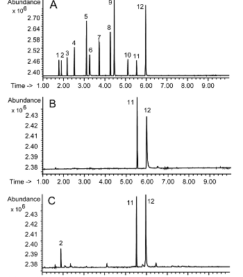
Fig. 1A shows the gas chromatogram of a standard mixture of stimulants, analyzed using a nitrogen- and phosphorus-specific flame ionization detector (NP-FID). Fig. 1B demonstrates a typical chromatogram of a urine extract, showing only caffeine (11), which is detectable in almost all urine samples, and an internal standard (12) (N-dodecyl-N,N-diisopropylamine). A positive amphetamine case is shown in Fig. 1C, where a distinct signal (2) with the retention time of amphetamine (see Fig. 1A with signal 2) is recorded.
GC/NP-FID chromatogram for unambiguous identification?
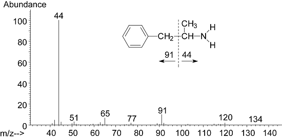
The GC/NP-FID chromatogram is not sufficient for the unambiguous identification of amphetamine (2) in Fig. 1. Mass spectrometric identification must be performed here. Fig. 2 shows the mass spectrum of amphetamine following electron impact ionization at 70 eV, wherein the intensity of the molecular ion m/z 135 (not visible) is significantly lower than 1% of the base peak. Following alpha-cleavage to nitrogen (Fig. 2), the spectrum is dominated by the fragment m/z 44 (base peak).
References
[1] Donike M, Jaenicke L, Stratmann D, Hollmann W: Gas-chromatographischer Nachweis von stickstoffahltigen Pharmaka in wässrigen Lösungen mit dem Stickstoffdetektor. J Chromatogr, 52 (1970) 237.