Hair analysis

Detection of Doping Substances in Human Hair: The active substances relevant to doping analysis are generally detected in urine. This sample has the advantage that, unlike blood collection, for example, providing a urine sample does not involve an invasive procedure. Furthermore, most substances are concentrated by the kidneys and are therefore present in higher concentrations in the urine.
Nevertheless, the detection of doping agents in urine is time-limited, meaning that if the substances are discontinued in time, detection is no longer possible. The analysis of hair samples offers entirely different detection windows.
After a medication is taken, the active ingredient enters the hair follicle via the bloodstream and grows through the scalp along with the hair shaft. Embedded in the hair’s protein structure, the substances are protected from further breakdown by enzymes and are effectively preserved until analysis.
Defined sample preparation procedure as a prerequisite
Before the hair sample can be analyzed, the substances of interest must be isolated and specially prepared. These steps are collectively referred to as the sample preparation procedure and serve to make the analyte (substance of interest) available in detectable quantities for the measurement method in the first place.
The information obtained from the analytical data allows for the unambiguous identification of the administered substance. By examining the hair in sections, the timing and duration of administration can be approximately reconstructed, taking into account individual hair growth rates. A hair analysis makes it possible to distinguish between chronic abuse and a single medically indicated administration, since a single dose of a medication does not lead to significant accumulation in the hair.
In certain cases, hair analysis also allows conclusions to be drawn about the administered parent compound. For example, unlike urine analysis, the ingestion of steroid esters can be detected in hair.
A disadvantage of this method lies in the melanin-dependent concentration of the substance, which means that detection of a substance may take longer in dark-haired athletes than in light-haired ones. Certain hair treatment methods, such as frequent perming, hair bleaching or dyeing, as well as incomplete hair lysis during sample preparation, can influence the results of a hair analysis.
Hair
root: Hair is formed in the hair root, where a specific protein structure called α-keratin is produced. The hair root is supplied with the necessary building blocks (including amino acids) via the bloodstream. As a result, almost all substances present in the blood can come into contact with the hair, including exogenous (externally supplied) substances. In addition to substance-specific properties such as molecular size, lipophilicity, and basicity, factors affecting the environment at the hair root—such as the pH gradient and concentration gradient of the substance in question—are also crucial for entry into the matrix cells of the hair roots and thus for incorporation into the hair shaft. Melanin, the pigment responsible for hair color, plays a key role in the transition from blood to hair. Melanin appears to act as a carrier, whereby the substance binds to the polyanionic structure of melanin and is transported through the membrane.
Hair follicles: Schematic representation of hair follicle morphology
The endogenous or physiological uptake discussed here must be fundamentally distinguished from exogenous uptake. In exogenous uptake, the foreign substance enters the hair from the environment. As a rule, such exogenous contaminants can be removed through washing steps using organic solvents and detergents. Many pharmacological substances are metabolized by the body and enter the hair as characteristic metabolites. Their detection is considered an important criterion for distinguishing between endogenous uptake and exogenous contamination.
Sample
Preparation Procedure
To ensure reproducible results, samples are collected from the occipital protuberance (back of the head) directly at the scalp. The hair strands, approximately 5 mm thick, are secured with adhesive tape or string to prevent slipping and stored in aluminum foil or glass containers until analysis. The hair sample obtained in this way must be prepared for the actual measurement through several chemical steps. First, external contaminants on the hair shaft must be removed using special reagents, such as methanol, dichloromethane, and lauryl sulfate solutions, so that only the portion of the substance that has entered the interior of the hair via endogenous uptake through the bloodstream is detected. To make this portion of the substance accessible for measurement, it must be released from the hair, i.e., the hair’s protein structure must be broken down.
Broadly speaking, there are two types of hair digestion methods:
1. Chemical/enzymatic and 2. Mechanical digestion methods. Certain chemicals (guanidine, and in some cases urea) have a denaturing effect on proteins. The addition of dithiothreitol reduces the numerous disulfide bonds in α-keratin and further destabilizes the protein.
Homogeneous solubilization (complete dissolution) of the hair can be achieved using strong acids or bases in combination with heat. Such methods have the disadvantage that they can also attack the analyte. Examples include the hydrolysis of steroid esters and the conversion of cocaine to benzoylecgonine, a natural metabolite of cocaine. As a result, the amount of benzoylecgonine produced by the body’s own enzyme system following cocaine consumption can no longer be verified. A gentler digestion can be achieved through enzymatic methods in which specific proteases cleave the peptide bonds within the protein molecule. The use of ball mills, in which the hair is mechanically pulverized without destroying the analyte, has also proven effective.
Extraction step following hair digestion
After hair digestion, an extraction step with a suitable organic solvent (methanol, ether) follows to isolate the target substance from the hair hydrolysate. The extraction yield for the analyte should be as high as possible and for interfering co-substances as low as possible, resulting in optimal detection sensitivity. In subsequent processing steps, the sample is dried, derivatized, and finally analyzed using GC/MS.
Substance
Concentration Concentrations of selected foreign substances in hair (literature data)
| Substance | Concentration in ng/mg | Substance | Concentration in ng/mg |
| Testosterone decanoate | up to 15 | Codeine | 0.1 to 3 |
| Nandrolone | up to 5 | Morphine | 0.1 to 38 |
| Amphetamine | 1 to 13 | Nicotine | 0.9 to 38 |
| Clenbuterol* | 0.005 to 0.25 | Secobarbital | 21 to 59 |
| Cocaine | 0.1 to 6 | Phencyclidine | 0.5 to 2 |
| Benzoylecgonine | 1 to 4 | Cannabinoids | 0.2 to 3 |
In the GC/MS method (gas chromatography/mass spectrometry), the isolated substances are bombarded with electrons following a preliminary gas chromatographic separation. In the process, the compound breaks down into characteristic molecular fragments that yield a substance-specific mass spectrum, thereby ensuring the unambiguous identification of the substance.
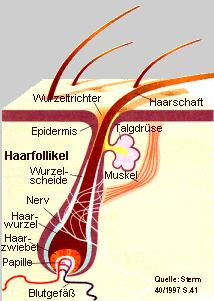
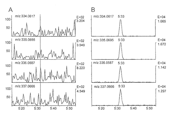
Figure 1B shows a Selected Ion Monitoring (SIM) chromatogram recorded following the analysis of a hair sample collected two months after the end of treatment. The sample was obtained from a patient who had taken clenbuterol for 35 days at a dosage of 3 × 20 µg/day.
The chromatogram contains 4 characteristic ion peaks at the retention time of clenbuterol. The signal intensities correspond to a concentration of approximately 90 ng clenbuterol per gram of hair. For comparison, Figure 1A shows a blank hair sample from the same patient, which was, however, collected prior to therapy.
Clenbuterol
Clenbuterol belongs to the group of β2-agonists and can be regarded as a synthetic derivative of naturally occurring catecholamines.
Clenbuterol (e.g., Spiropent) is approved in human medicine as a bronchodilator and tocolytic (contraction inhibitor). In animal studies, an increased rate of lipolysis and anabolic effects have been demonstrated at doses 5 to 10 times higher than the therapeutic dosage. These effects have led to the illegal use of clenbuterol in animal fattening to increase meat production.
In humans, the misuse of clenbuterol is intended to increase muscle mass while simultaneously minimizing body fat. Therefore, clenbuterol is used by athletes in strength-based disciplines such as bodybuilding, track and field, and weightlifting. Due to clenbuterol’s short half-life in the body, it can only be detected in urine for a short period (24 hours to a few days).
Fig. 2: Chemical structure of clenbuterol
References
Machnik, M., Schänzer, W.: Nachweis von Dopingsubstanzen im Humanhaar. F.I.T., Wissenschaftsmagazin der Deutschen Sporthochschule Köln, 1 (2001) 12 15.
download des Artikels
Institute of Biochemistry at the German Sport University Cologne, December 12, 2001
